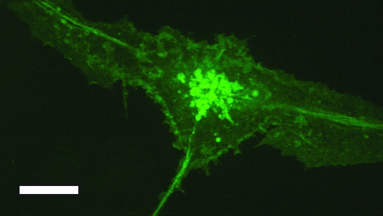
Figure 1. Spinning disk confocal image of class II MHC-GFP expressing primary bone marrow derived DC. Note that most of the GFP signal is found intracellularly. Endolysosomal tubes are present. Scale bar represents 5 µm.

Figure 1. Spinning disk confocal image of class II MHC-GFP expressing primary bone marrow derived DC. Note that most of the GFP signal is found intracellularly. Endolysosomal tubes are present. Scale bar represents 5 µm.