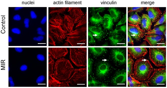
Figure 3. Effect of MIR exposure on the actin filaments and focal adhesions of A549 cells.
Cells were seeded onto glass coverslips in 12-well plates, exposed to MIR for 48 hours, fixed for staining and visualized by fluorescence microscopy. Actin filaments were tagged with rhodamine-labeled phalloidin (red), vinculin was labeled with mouse anti-vinculin antibody and the corresponding FITC– conjugated secondary anti-mouse IgG antibody (green), and nuclei were stained with DAPI (blue). Scale bar represents 10 µm. Arrows indicate the position of vinculin.

