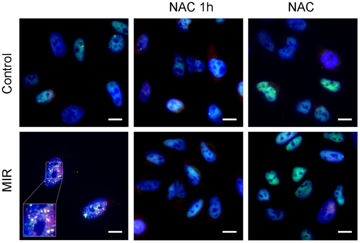
Figure 6. Effect of MIR exposure on DNA double strain breaks in A549 cells.
Cells were seeded onto the glass coverslip in 12-well plate, exposure by MIR for 48 hours in the presence or absence of 10 mM N-Acetyl-Cysteine (NAC). Cells were treated with NAC for 1 h prior to MIR exposure (NAC 1 h) or cotreated throughout the exposure for 48 h (NAC). Cells were fixed for staining and visualized by fluorescence microscopy. 53BP1 was labeled with rabbit anti-53BP1 antibody and corresponded FITC–conjugated anti-rabbit IgG antibody (green), γ-H2AX was labeled with mouse anti-γ-H2AX antibody following corresponded PE–conjugated anti-mouse IgG antibody (red), and nuclei were labeled with DAPI (blue). Scale bar represents 10 µm.

