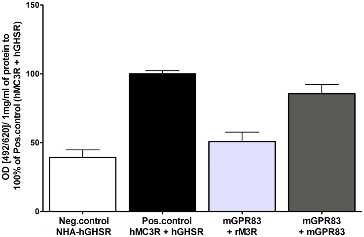
Figure 4. Homodimerization of mGPR83.
Dimerization studies were performed using sandwich ELISA. COS-7 cells were transiently transfected. As negative control serves the NHA-hGHSR (white column) and as positive control the co-transfection of NHA-tagged hMC3R and FLAG-tagged hGHSR (black column, [28]). The light grey column represents the average of the HA- respectively FLAG-tagged mGPR83 in combination with the correspondent tagged rM3R. The dark grey column represents co-transfection of HA-tagged mGPR83 and FLAG-tagged mGPR83. Dimerization was measured via the HA epitope. The mean absorption (492 nm/620 nm) is calculated per 1 mg/ml of protein and shown as percentage of the hMC3R/hGHSR heterodimer (absorption (492/620)/ 1mg/ml of protein: 0.3 ± 0.04). Data were assessed from 3 independent experiments, each performed in triplicates and represent mean ± SEM.

