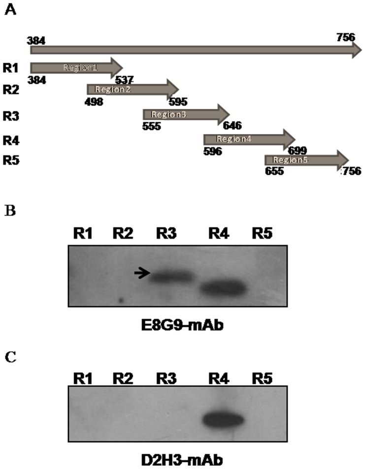
Figure 4. Epitope mapping of E8G9.
(A) Schematic representation of different fragments of HCV E2 protein used for epitope mapping. (B) Western blot analysis of the recombinant proteins from five regions of E2 (region 3 specific for E8G9 is indicated using an arrow). (R1–R5 denote different regions).

