
Figure 3. Other examples of TDP-43 pathology.
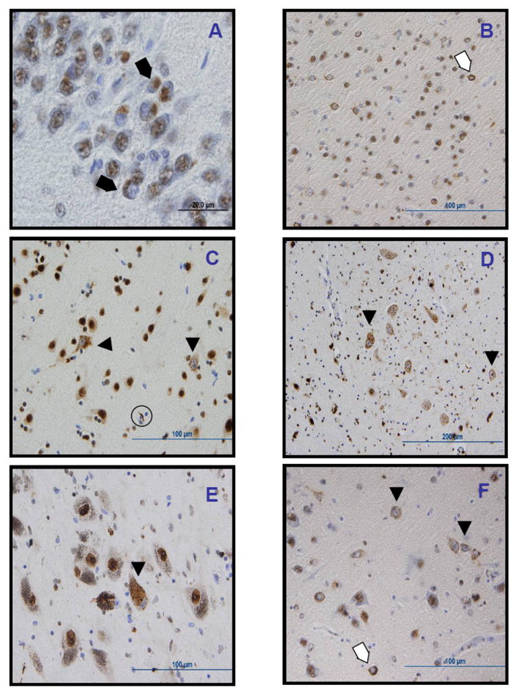
Other TDP-43 positive lesions included dense round inclusions in the dentate gyrus of the hippocampus in case III-4 (A); ring-like dense cytoplasmic inclusions (thin arrows) and diffuse stippled cytoplasmic staining with nuclear clearing or “pre-inclusions” (arrowheads) in the entorhinal cortex (B), temporal cortex (C), hyppoglossal nucleus (D), substantia nigra (E), and amygdala (F), as well as a glial inclusions (thick arrow, C). All images obtained from case III-4, except for C, from case II-7.