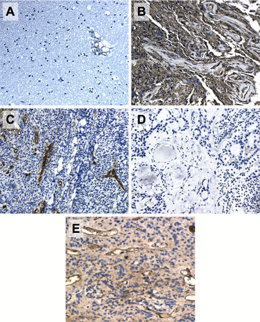
Fig. 2.
Representative immunohistochemical images of HLA Class I in normal brain (a), glioblastoma with known positive expression (b, positive control), medulloblastoma (c, negative control), ependymoma with negative immunoreactivity (d), ependymoma with positive immunoreactivity (e). Images at original magnification (100×) are shown

