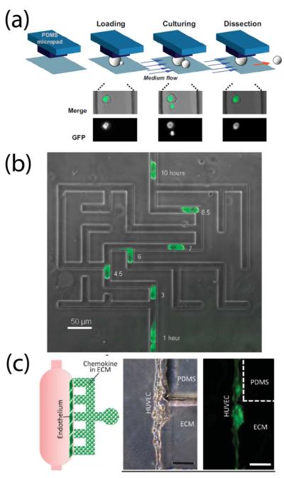
Figure 5.
Control of the cellular microenvironment. μTAS technology enabled new studies in a variety of biological systems. (a) Selective release and tracking of newly-budded yeast daughter cells across multiple generations controlled by microfluidic flow. Reprinted with permission from ref 147. Copyright 2012 National Academy of Sciences. (b) A microfluidic maze established tunable EGF gradients on the cellular level to study epithelial cell migration. From ref 153. Reproduced with permission of the Royal Society of Chemistry. (c) Chemokine-induced adenoid cystic carcinoma intravasation through a mock endothelial cell monolayer in microfluidic-based device. Scale bar, 200 μm. From ref 159. Adapted by permission from the Royal Society of Chemistry.

