Figure 6.

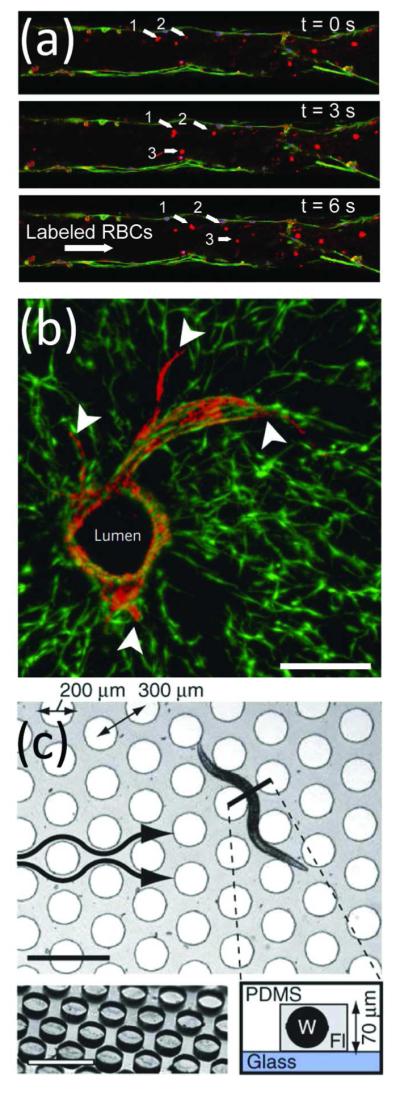
Organs and organisms-on-chip. Microfluidics allowed controlled studies of (a,b) cell-cell interactions and (c) whole organisms. (a) Time-lapse images of a red blood cells flowing through a capillary network developed from endothelial cells cultured in a microfluidic channel. From ref 165. Reproduced with permission of The Royal Society of Chemistry. (b) Sprouting of mCherry-expressing endothelial cells (arrowheads) from a central lumen was demonstrated within a co-culture of 10T1/2 cells expressing enhanced green fluorescent protein. Scale bar, 200 μm. Adapted by permission from Macmillan Publishers Ltd: Nature Materials, ref 170, copyright 2012. (c) A microfluidic device for examining behavioral responses of C. elegans to chemical changes. Scale bars, 500 μm. Adapted by permission from Macmillan Publishers Ltd: Nature Methods, ref 180, copyright 2012.
