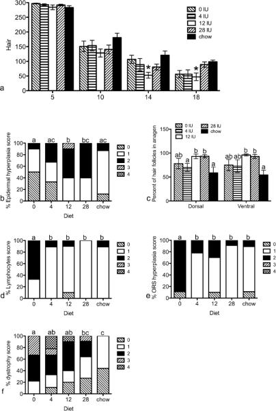
Figure 3. Progression of AA was altered by dietary vitamin A.
Mice were fed purified diets containing 0, 4, 12, or 28 IU dietary vitamin A/g diet, or a control chow diet starting two weeks prior to grafting and sacrificed 5, 10, 15 (b, c), or 20 (d, e, f) weeks post grafting. Ventral hair loss (a) was measured weekly with 300 representing a full coat of hair. Data are shown as mean ±SEM. H&E slides were scored by JP Sundberg on a scale of 0–4. Data are shown as percent of each score for epidermal hyperplasia (b), anagen (c), lymphocytes (d), outer root sheath hyperplasia (e), and follicular dystrophy (f). n=8–11. *=significantly different from chow fed mice, p<0.05, different letters are significantly different, p<0.05.

