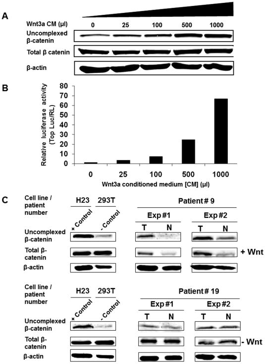
Figure 1.
Uncomplexed b-catenin level is a valid marker for Wnt activation in non-small cell lung cancer (NSCLCs). 293T cells transduced with TOP-luciferase and renila luciferase lentiviruses were treated for 48 hours with increasing concentration of Wnt3a conditioned medium. (A) Glutathione S-transferase (GST)-E cadherin capture assay as described in the Methods was performed on the treated cells Total cell lysates (20 μg) and the GST-E-cadherin precipitates from 800 μg of cell lysate were subjected to immunoblot analysis with a mAb directed against β-catenin. (B) TCF reporter luciferase activity was performed on 1:10 of cell volume from the same Wnt3a conditioned medium treated cells as described in Methods. Luciferase reporter activity was calculated by dividing the TOP-luciferase by renila luciferase (RL). (C) β-catenin capture assay performed with two representative human paired NSCLC and normal lung tissues in two independent experiments. Total cell lysates (800 μg) were subjected to GST-E cadherin capture assay as described in Figure 1A. T denotes tumor specimen and N denotes normal lung tissue from the same patient. H23, a Wnt autocrine activated NSCLC line, and 293T, an immortalized human embryonic kidney line, were used as positive and negative controls, respectively.

