
Figure 1. Active site of PchB in the open and closed wildtype and closed K42A-PchB structures.
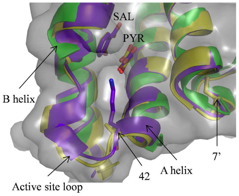
Overlay of the wildtype closed (PDB: 3REM, purple), wildtype open (PDB: 2H9C, green) and K42A –PchB (PDB: 3HGX, yellow) structures. The open structure, solved in the absence of products, has no electron density for the active site loop, which connects the A and B helices and contains a single turn of helix when ordered in the closed structures. The amino acid at the 42 position, which is the first residue of the active site loop, is shown as sticks, as is the amino acid at the 7 position from the opposing monomer (7′). The salicylate and pyruvate from the closed structures are also shown as sticks, all colored like the cartoon. The solvent accessible surface (grey) for the K42A structure shows a hole leading to the active site, highlighting the location that a chemical rescue agent must bind and be organized. Figure was generated with PyMOL (52).