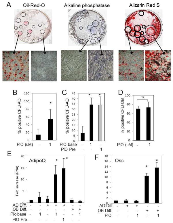
Fig. 4. Pioglitazone alters adipocyte CFU but has no effect on osteoblast CFU from human bone marrow.
Whole bone marrow was diluted 1:20 in DMEM and colonies allowed to expand for twenty-one days. Cells were then treated with AD or OB differentiation medium and +/− PIO (1μM) as indicated. Colonies were treated with adipocyte (CFU-AD) or osteoblast (CFU-OB) differentiation medium and stained for adipocyte positive colonies with Oil-red-O or osteoblast positive colonies with either Alizarin Red S or NBT/BCIP (alkaline phosphatase activity). (A) Representative wells from a 6-well plate demonstrate both positive and negative colonies for the different stains. (B) The CFU-AD assay was performed in the presence or absence of 1μM PIO and positive colonies counted and expressed as a ratio relative to the total number of colonies. (mean of 7 patients). (C) Colonies were pretreated for 5–14 days with 1μM PIO prior to the initiation of adipogenesis and the percent of positive colonies relative counted and expressed as a relative ratio to the total number of colonies. (mean of 7 patients). (D) The CFU-OB assay was performed in the presence or absence of 1μM PIO and positive colonies counted and expressed as a ratio relative to the total number of colonies. (mean of 7 patients). Parallel plates were harvested for RNA analyses and analyzed for adipocyte marker genes, adiponectin shown (E), and osteoblast marker genes, osteocalcin shown (F) by qRT-PCR (Rep. of 7 patients). * p < 0.05. Results are presented as mean+/−SEM. ns:not significant

