
Figure 1.
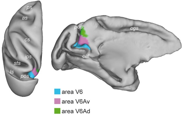
Brain location of macaque area V6. Dorsal (left) and medial (right) views of the surface-based 3D reconstructions of the ATLAS brain of the macaque obtained by CARET (Computerized Anatomical Reconstruction and Editing Toolkit, http://brainvis.wustl.edu/wiki/index.php/Caret:About) (Van Essen et al., 2001) showing the extent of area V6 (light blue) on the left hemisphere. pos, parieto-occipital sulcus; cal, calcarine sulcus; cgs, cingulate sulcus; ips, intraparietal sulcus; sts, superior temporal sulcus; ls, lunate sulcus; cs, central sulcus; as, arcuate sulcus; ps, principal sulcus.