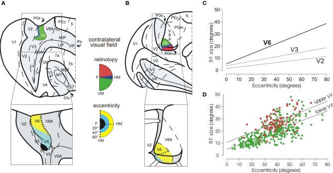
Figure 2.
Visual topography and receptive field properties of macaque area V6. (A) Dorsal view of caudal half of right hemisphere (and, below, enlargement of the parieto-occipital region) with the parieto-occipital (POs), lunate (Ls), and intraparietal (IPs) sulci shown opened to reveal the cortex buried within them (dark gray area). (B) Medial view of the caudal half of a the left hemisphere (and, below, enlargement of the parieto-occipital region), with the medial parieto-occipital sulcus (POs) opened. Area V6 is shown in color, according to the part of visual field it represents (conventions reported between A and B). Note that V6 represents point-to-point the entire contralateral visual field, with an emphasis in the representation of the peripheral visual field. Triangles and crosses indicate the representation of the horizontal (HM) and vertical (VM) meridians of area V6, respectively; the F, the center of gaze. Dashed lines are the borders between different cortical areas. PEc, 5, MIP, LIP, VIP, 7a, 7b, MT, MST, V4, V4T, FST, PGm: areas functionally or anatomically identified in the posterior part of the cerebral hemisphere. Modified from Pitzalis et al. (2006). (C) Regression plots of receptive-field size (square root of area) against eccentricity for cells recorded in areas V2, V3, and V6. Receptive-field size increases with eccentricity in all visual areas. In area V6, receptive fields are larger than in V2 and V3 at any eccentricity. (D) Dual regression plot of RF size against eccentricity for RF in the upper (red circles) or lower (green circles) visual fields (VF), respectively. Modified from Galletti et al. (1999a). It is evident that at any eccentricity, RFs are bigger in the upper VF with respect to the lower one.

