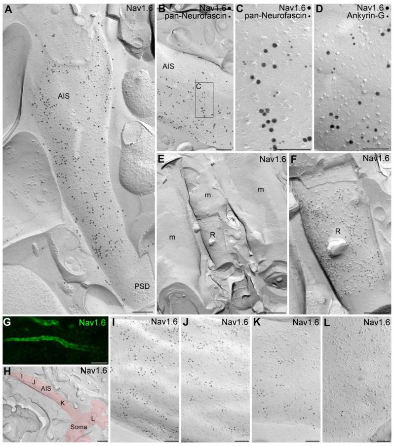
Fig. 3.
High resolution immunogold localization of the Nav1.6 subunit in AISs and nodes of Ranvier of CA1 PCs. (A) A high density of gold particles labeling the Nav1.6 subunit is found on the P-face of an AIS in the stratum pyramidale. Note that the immunogold particles avoided the area of the postsynaptic density (PSD) of an axo-axonic synapse. (B) An AIS is co-labeled for the Nav1.6 subunit (15 nm gold) and the AIS marker pan-Neurofascin (10 nm gold). (C) A high magnification view of the boxed area shown in B. (D) High magnification image of the P-face of an AIS labeled for the Nav1.6 subunit (15 nm gold) and Ankyrin-G (10 nm gold). (E) A low magnification image shows a node of Ranvier (R) of a myelinated axon (m) in the alveus. (F) A high magnification view of the node of Ranvier shown in E. The P-face of the node of Ranvier membrane contains a high density of gold particles labeling the Nav1.6 subunit. Note the lack of labeling over the myelin (E and F). (G) A gradual increase in the intensity of Nav1.6 immunofluorescence is found along the proximo-distal axis of CA1 PC AISs. (H) A low magnification image of a replica shows a fragment of a somatic plasma membrane and an emerging AIS (red). (I-L) High magnification images of the boxed areas shown in panel H. Images taken of the soma (L) and of the AIS at various distances from the soma (I-K) demonstrate an increase in the density of gold particles labeling the Nav1.6 subunit along the proximo-distal axis of the AIS (I). Scale bars: A, B, F, 200 nm; C, D, I-L, 100 nm; E, 500 nm; G, 5 μm; H, 1 μm.

