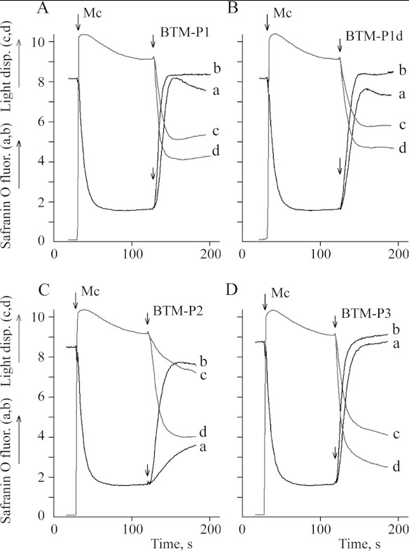
Figure 1. Influence of the peptides derived from the Cry11Bb protoxin on the inner membrane potential of rat liver mitochondria monitored by safranin O fluorescence (a,b), and on mitochondrial swelling monitored by light dispersion (c,d).
Mitochondria (0.5 mg/ml protein) were added to the KNPE medium supplemented with 2.5 mM succinate and 10 μM safranin O; (a,c) 0.5 μM peptide; (b,d) 1.0 μM peptide.

