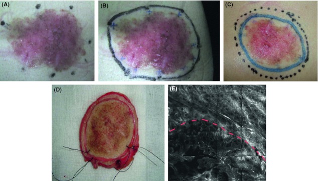
Figure 1.

Reflectance confocal microscopy imaging protocol and surgical excision. (A) Foci for reflectance confocal microscopy (RCM) examination were selected. (B) The lesion's border was refined (blue cross). (C) An extra 5-mm margin of normal skin was excised (dotted line). (D) The gross skin specimen was further sectioned along the boundaries inscribed using RCM. (E) RCM mosaic image of the margin. The upper half part of the image showed the normal reticulated meshwork pattern, while the lower part showed the tumor islands of elongated strands.
