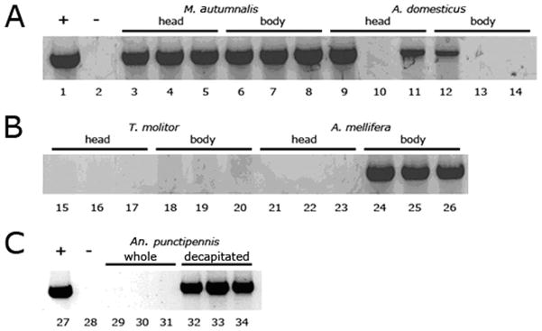
Fig. 4.

Survey for PCR inhibitors from various insects. Positive control DNA (1 μl; lanes 1 and 27) was combined with 8-μl template DNA from various extractions (lanes 3–34). Lanes with reduced PCR product, relative to the positive control show evidence for an inhibitor. Panels A and B show lanes from the same gel, whereas Panel C is from a separate gel. Lanes 2 and 28 are negative controls without DNA. Lanes 3–5 show M. autumnalis head DNA and 6–8 show M. autumnalis decapitated, whole body DNA. Lanes 9–11 show A. domesticus head DNA and 12–14 show A. domesticus decapitated, whole body DNA. Lanes 15–17 show T. molitor head DNA and 18–20 contain T. molitor decapitated, whole body DNA. Lanes 21–23 show A. mellifera head DNA and 24–26 show A. mellifera decapitated, whole body DNA. Lanes 29–30 are An. punctipennis whole mosquitoes and 32–34 are decapitated An. punctipennis mosquitoes.
