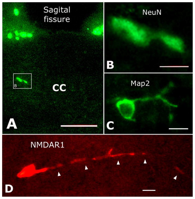
Fig. 4.
Double immunostaining with anti-NMDAR1 and anti-NeuN or anti-MAP2, but no double-labeled structures were observed at all. A: NeuN-positive neurons located in the CC. B: High magnification of the boxed area in A. C: MAP2-labeled neuron in the CC. D: NMDAR1-immunostained cell with a long process (arrowheads). Scale bars = 50 μm in A; 15 μm in B–D.

