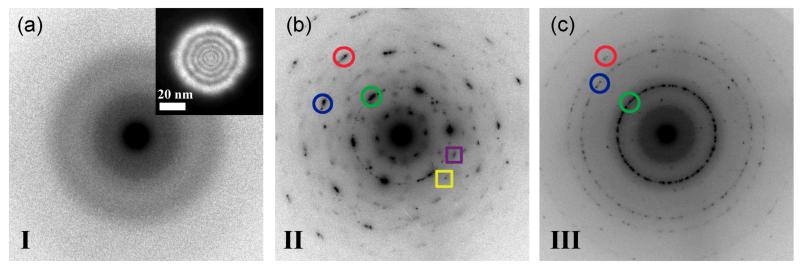
Figure 3.
Nano-area diffraction images taken using a ~50 nm parallel electron probe as shown in the inset. (a) NED image of the Al2O3 support prior to pore formation shows that the membrane is amorphous (b) NED pattern obtained by placing parallel electron probe over pore located in stage II of Figure 2b. Distinct spot reflections marked with circles are observed corresponding to formation of γ and α-Al2O3 nanocrystallites. Additional reflections marked by squares are from δ and κ phases (c) NED pattern obtained by placing parallel electron probe over pore located in stage III of Figure 2b. Continuous rings are observed confirming that a polycrystalline material with preferred phases (γ and α) is formed by prolonged exposure to the convergent beam.

