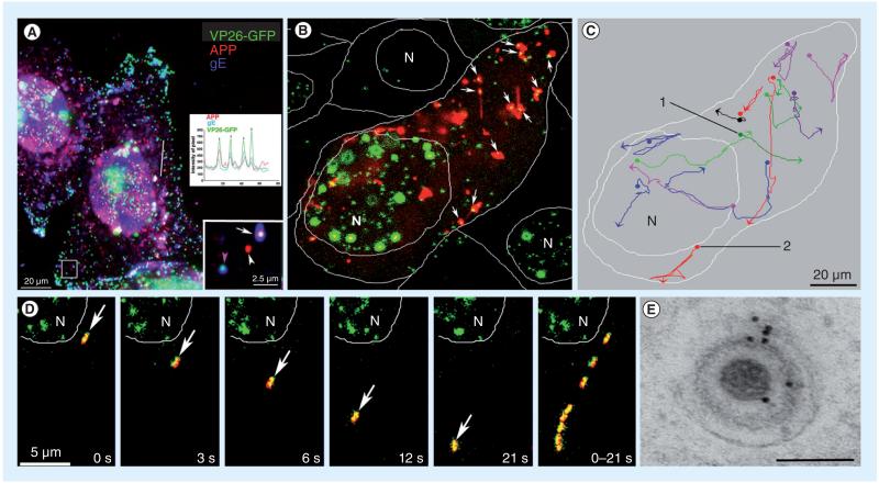
Figure 2. HSV-1 interacts with cellular amyloid precursor protein during egress.
(A) VP26-GFP HSV-1 (green) particles colocalize with APP (red) during egress by immunofluorescence after synchronous infection with VP26-GFP HSV-1 (green) and fixation at 7 h postinfection. Examples of two infected cells also stained by immunofluorescence for gE (blue). Lower right inset shows a higher magnification of the boxed region. In this inset, one VP26-GFP-labeled particle displays all three labels (gE, APP and VP26), another has only APP (white arrowhead) or only gE (pink arrowhead). Such single labels demonstrate that colocalization is not due to bleed-through from other fluorescent channels. The inset graph shows intensity profiles along the line drawn across the right-hand cell. Superimposed peaks in the graph for the different colors are indicated by vertical arrows. (B) The first frame of a video sequence showing the initial positions of HSV capsids (VP26-GFP, green) and cellular APP-labeled with red fluorescent protein (APP-monomeric red fluorescent protein, red) in an infected cell at 7–9 h postinfection from a 3-s time-lapse 900-s (15 min) video. Double-labeled particles appear yellow (arrows). Many capsids (64%) colocalize with APP compartments in this frame. Capsids travel with APP vesicles, and sometimes join and separate from them. Positions of the N and of the cell boundaries are delineated by white lines. (C) The tracks of selected VP26-GFP HSV particles that move with the APP-monomeric red fluorescent protein label. Each trace has been assigned a different pseudocolor. Beginnings and ends of movements are indicated by dots and arrowheads, respectively. (D) An example of a double-labeled HSV-APP particle that moves away from the nucleus (arrow). In the last panel eight frames are superimposed to demonstrate the particle’s trajectory. (E) Immunogold-labeled electron micrograph of an HSV-1 particle inside an infected cell cytoplasm probed with anti-C-APP followed by protein-A labeled with 10 nm gold. Gold particles decorate both cellular and viral membranes surrounding capsids.
Scale bar = 100 nm.
APP: Amyloid precursor protein; GFP: Green fluorescent protein; N: Nuclei.
Modified with permission from [7]. Supporting videos for these images can be found on the PLoS One journal website linked to the online publication.

