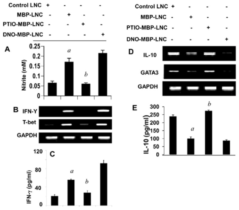
Figure 2. Differential regulation of T-bet/IFN-γ and GATA3/IL-10 by NO.

Lymph node cells (LNC) isolated from MBP-immunized female SJL/J mice were stimulated with 50 μg/ml MBP in the presence or absence of PTIO (50 μM) and DETA-NONOate (DNO; 100 μM), respectively. After 24 h of stimulation, the concentration of nitrite was measured in supernatants using ‘Griess’ reagent (A) and the mRNA expression of T-bet and IFN-γ (B) and GATA3 and IL-10 (D) was monitored in cells by semi-quantitative RT-PCR. After 72 h of stimulation, supernatants were assayed for either IFN-γ (C) or IL-10 (E) by ELISA. Results are mean + SD of three different experiments. ap<0.001 vs control; bp<0.001 vs MBP.
