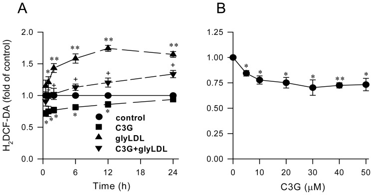
Figure 1.
Effect of C3G on redox status in EC. PAEC were treated with vehicle (control), 30 μM C3G, 100 μg/mL of glyLDL or C3G + glyLDL for 0.5–24 h (A) or with 0–50 μM C3G for 30 min (B). Redox status was assessed by measuring fluorescence intensity in cells at 485/530 nm (excitation/emission) using a fluorescence microplate reader. Values were expressed in mean ± SD in fold of control (n = 3 independent experiments). *, **: p < 0.05 or 0.01 versus control; +: p < 0.05 versus glyLDL.

