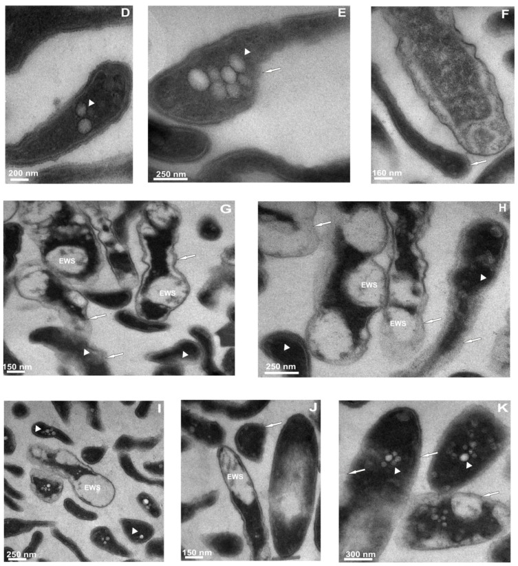
Figure 4.
The influence of defensin and apolipophorin III isolated from G. mellonella hemolymph on the ultrastructure of L. dumoffii cells. The cells grown on the non-supplemented (A–C,G,H) and choline-supplemented (D–F,I–K) medium were incubated in the presence of apoLp-III (A–F) or Galleria defensin (G–K). Then the cells were prepared for TEM analysis as described in the Experimental Section. (A,B) cells showing condensed cytoplasm, regions with decreased electron density and the presence of vacuoles (arrowhead); (C) a fragment of the bacterium with a group of visible vacuoles and dark, dense cytoplasm; (D,E) cells with vacuolization features (arrowheads) and cell envelope damage (arrow); (F) bacteria with cell wall distortion (arrow) and a widened periplasmatic space; (G) cells with electron-white spaces (EWS), membrane deterioration (arrows), and condensed content (arrowheads); (H) irreversible cell wall damage visible in bacteria (arrows) together with dense areas of cytoplasm or entire cytoplasm of the whole cells (arrowheads); (I) bacteria demonstrating loss of cell wall integrity, vacuolization of cytoplasm (arrowheads), cell shrinkage; and (J,K) cells showing cell wall damage (arrows), electron-white spaces (EWS), presence of small vacuoles (arrowheads).


