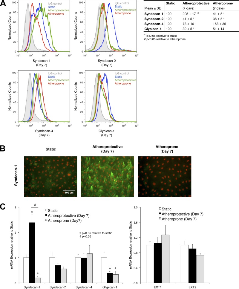
Fig. 4.
Prolonged exposure to atheroprotective flow induces expression of syndecan-1 on the apical surface of endothelial cells. A: flow cytometry histograms of syndecan-1, -2, and -4 and glypican-1 surface expression on cells cultures under static condition or exposed to atheroprotective or atheroprone shear stress waveform for 7 days and quantitative analysis of flow cytometry data from 3 independent experiments. Values are means ± SE (n = 3). B: representative immunostaining images of syndecan-1 in cells cultured under static condition or exposed to atheroprotective or atheroprone flow for 7 days. Syndecan-1 is shown in green, and nucleus (DAPI) is shown in red. C: mRNA expression of heparan sulfate protein carriers (left) and key enzymes specifically responsible for heparan sulfate chain biosynthesis (right). Values are means ± SE (n = 3).

