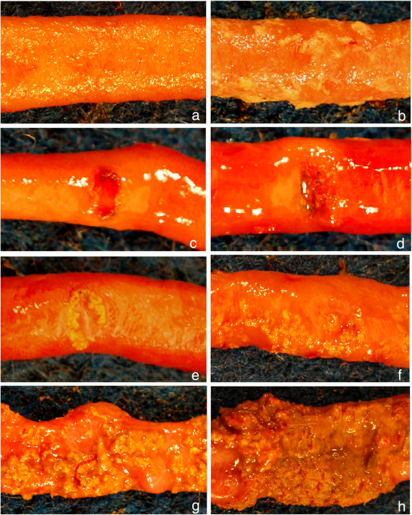
Figure 2.
Different lesions of necrotic enteritis in chickens, used to illustrate the scoring system (Table 2 ).a: Necrotic enteritis score 0, everted jejunal segment. No gross lesions are present. b: Necrotic enteritis score 1, everted jejunal segment. There are no obvious ulcers in the mucosa, but the entire mucosal surface is covered with a layer of loosely adherent fibrin. c: Necrotic enteritis score 2–4, everted jejunal segment. There is an excavated ulcer of the mucosa with acute, bright red hemorrhage within the ulcer bed and scant crusting of fibrin around the periphery. d: Necrotic enteritis score 2–4, everted jejunal segment. There is an excavated ulcer of the mucosa with dark green-black pigment within the ulcer bed and scant crusting of fibrin over the surface. e-f: Necrotic enteritis score 2–4, everted jejunal segments. There are excavated ulcers of the mucosae, the periphery of which are covered by thick, tightly-adherent layers of fibrin, necrotic tissue, and inflammatory cells. g-h: Necrotic enteritis score 5–6, everted jejunal segment. The mucosae are covered by large, confluent plaques of fibrin, necrotic tissue, and inflammatory cells (g) to the point where they extend over broad regions of the intestinal mucosa (h).

