
Figure 1. HFD induced oxidative stress, and decreased ATP levels in skeletal muscle and liver.
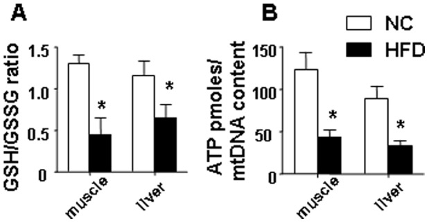
(A) the GSH/GSSG ratio was significantly decreased in both gastrocnemius muscle and liver samples isolated from mice fed a HFD. (B) a HFD reduced the ATP levels in both skeletal muscle and liver. ATP concentrations were determined using the luciferase-based ATP-assay, values were normalized to mtDNA content. The average results ± SE are shown. (*p<0.05 vs corresponding NC, n = 7–9 mice per group).