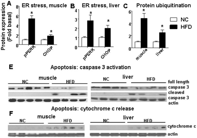
Figure 6. HFD induced markers of ER stress, protein degradation and increased apoptosis in skeletal muscle and liver.
pPERK/PERK and CHOP levels were increased after a HFD in both skeletal muscle (A) and liver (B). (C) HFD increased protein ubiquitination (a marker of both ubiquitin-proteasome and autophagy-induced protein degradation) in both skeletal muscle and liver. The average results ± SE are shown. (* p<0.05 vs corresponding NC, n = 6–9 mice per group). (E) Caspase 3 and cleaved caspase 3 antibodies were used to recognize full length (35 kD) caspase 3 and cleaved caspase 3 large fragment (17 kD), respectively. (F) Western blot of cytochrome c release into cytosol. Cytosolic fractions isolated from skeletal muscle and liver from NC/HFD fed mice are shown. Equal loading was confirmed using anti actin antibody (n = 4–5 mice per group).

