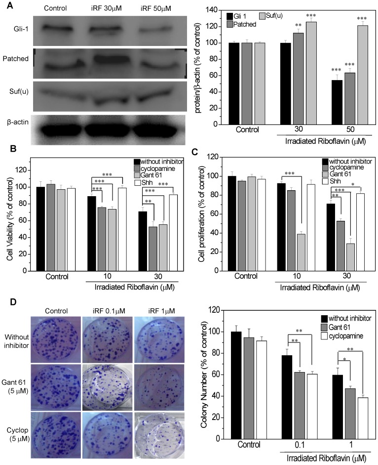
Figure 5. The effect of iRF on Hedgehog pathway.
(A) Western blotting analysis of the expression of GLI1, PTCH and SUFU in B16F10 treated with different concentrations of iRF for 24 h. (B) Cell viability (MTT assay) of B16F10 cells with or without pretreatment with 5 µM cyclopamine, 5 µM Gant61 and 0.5 µg/mL SHH for 6 h and following iRF treatment for 24 h. (C) Cell proliferation BrdU assay with or without HH modulator pretreatment followed by iRF treatment and (D) representative pictures of colony assay and the number of colonies of B16F10 cells treated for 10 days. The results are expressed as the mean ± SD and are representative of three independent experiments to western blotting and n = 9 for others experiments. p<0.05, p<0.01, p<0.001 versus control.

