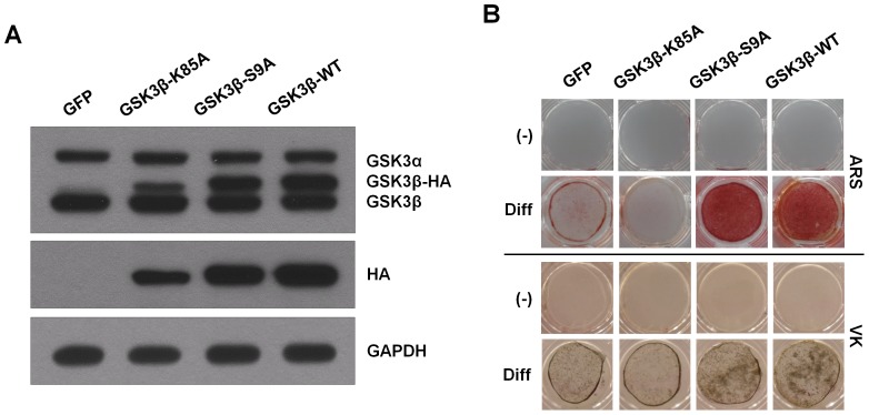
Figure 1. GSK3β activity regulates osteoblast differentiation in ADSCs.
(A) ADSCs were infected with either the retrovirus expressing GFP, catalytically inactive GSK3β (GSK3β-K85A), constitutively active GSK3β (GSK3β-S9A), or wild-type GSK3β (GSK3β-WT). After 48 hours, cells were harvested for immunoblot analysis for GSK3α/β expression using antibodies specific for GSK3α/β. It should be noted that due to the increased size of the HA-tagged GSK3β, the protein migrates at a higher molecular mass than that of endogenous GSK3β. The proper expression of transiently transfected HA-tagged GSK3β proteins was further confirmed by immunoblotting with anti-HA antibody. GAPDH served as a loading control. (B) Retroviral infected cells were cultured in osteogenic differentiation medium for 2 weeks. Enhanced matrix mineralization was visualized by Alizarin red S (upper, ARS) and von Kossa (lower, VK) staining. These results are representative of at least three independent experiments.

