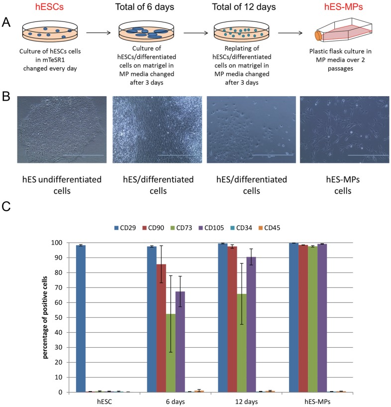
Figure 1. Derivation of hESC-MPs. A. Flow chart of experimental procedure for hESC-MPs derivation.
After culture on matrigel in mTESR1 media undifferentiated hESCs were shifted to MP specific media for 6 days. After 6 days the confluent culture of differentiated cells were split to 1/3 with dispase and replated on matrigel. After an extra 6 days of culture, differentiated cells were plated on coating-free feeder-free culture dishes. Cells were analyzed for MP markers expression at each step of the process. B. Phase contrast imaging of cell morphology modification during the derivation process of hESC-MPs. C. Chart representing flow cytometry analysis of mesenchymal progenitor markers CD29, CD90, CD73 and CD105 at the different time-points of our differentiation process.

