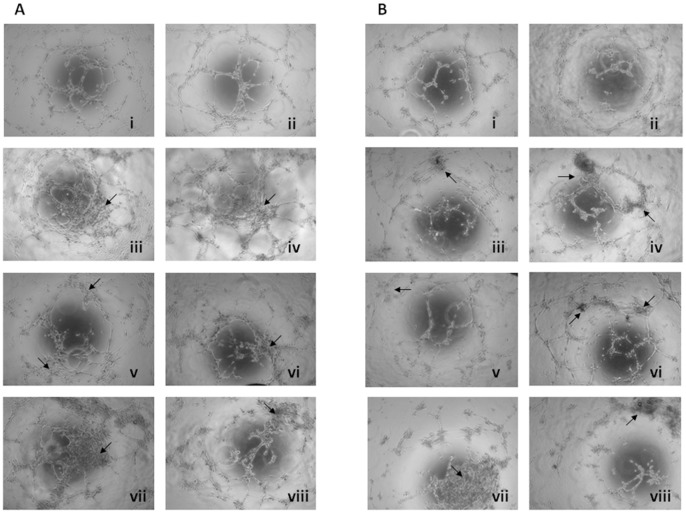
Figure 2. Effect of the MDS BM microenvironment on BMEC-1 and HMVEC-L tube formation.
BMEC-1 (A) and HMVEC-L (B) were seeded at a concentration of 8,000 cells per well of 96-well plate and incubated for 7 h at 37°C in 5% CO2. The endothelial tube formation was photographed at 5 h using a phase contrast inverted microscope. Each experiment was performed in duplicate. The pictures show the appearance of endothelial cell tubes on Matrigel® precoated plates in culture medium (i) and BM supernatant fluid from healthy control (ii), RA (iii), RARS (iv), 5q syndrome (v), RAEB (high-risk MDS) (vi) and RCMD (vii-viii) patients at 1∶10 dilution in culture medium. As the arrows show in the figure, the tube morphology was strikingly influenced by BM supernatant fluid from MDS (iii-viii) with respect to the controls (ii). The tubes originated after the incubation of BMEC-1 or HMVEC-L with the BM supernatant fluid from RCMD patients (vii-viii) were almost completely disrupted and formed closed capillary networks. MDS: myelodysplastic syndrome; BM: bone marrow; BMEC-1: bone marrow endothelial cells; HMVEC-L: lung-derived normal human microvascular endothelial cells; RA: refractory anemia; RARS: refractory anemia with ring sideroblast; RAEB refractory anemia with excess of blasts; RCMD: refractory cytopenia with multilineage dysplasia. (Controls n = 13; RA n = 5; RARS n = 6; 5q syndrome n = 2; RAEB n = 4; RCMD n = 7).

