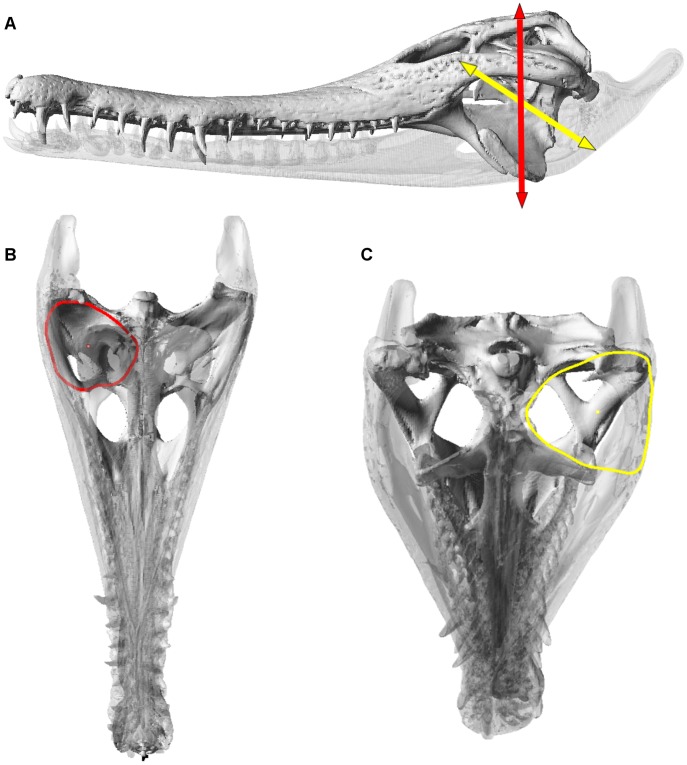
Figure 12. Reptile version of ‘dry skull method’ in a crocodile skull.
Skull of Mecistops cataphractus, showing: (A), temporal (red) and pterygoid (yellow) muscle vectors; temporal vector is oriented vertically with the skull aligned horizontally, pterygoid vector runs between a point that is half of the cranial height at the postorbital bar, to the ventral surface of the mandible directly below the jaw joint. (B), calculation of the cross sectional area (CSA) for the temporal muscles; the outline maps the extent of the adductor chamber defined from osteological boundaries, viewed normal to the relevant vector. (C), calculation of CSA for pterygoid muscles; the outline is drawn normal to the vector. Outlines in B and C also show centroids, used for calculation of inlevers (see Thomason [15], McHenry [29]).

