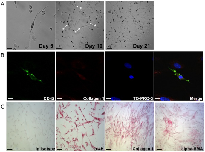
Figure 5. Characterization of bronchoalveolar lavage cells in culture.
A) Morphology of adherent bronchoalveolar lavage (BAL) cells at amplification when a positive culture occurred. From left to right: after 5 days (scale bar = 25 µm), at 10 days (scale bar = 250 µm) and at 21 days (scale bar = 250 µm). The arrows indicate typical adherent mesenchymal cells with high (*, fibrocyte phenotype) or low ratio (#, fibroblast phenotype) of cell length to cell width. B) Confocal microscopy analysis of fibrocytes (spindle-shaped cells co-expressing CD45 and collagen) in BAL cell culture at day 21. From left to right: CD45, collagen 1, thiazole orange protein 3 (TO-PRO-3) and merge of the three fluorescence channels. Scale bars = 15 µm. C) Representative immunocytochemical stain prepared from alveolar fibroblasts at passage 1. From left to right: Ig Isotype, α-propyl-4-hydroxylase (α-4H), collagen 1, α-smooth muscle actin (a-SMA). Scale bars = 100 µm.

