
Fig. 2.
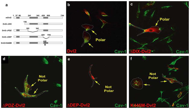
Dvl2 mutants disrupt endothelial cell polarity. a Diagram of HA-Dvl2 constructs in pCMV5 (HA-tags on the N-terminal of each construct are not depicted). Each of the functional domains have been individually deleted. In addition, one construct was generated through the introduction of a point mutation to yield the PCP-inhibitory K446M-Dvl2 construct. Caveolin-1 localization (b–f, green) in MPE cells expressing various HA-Dvl2 (red) constructs: b Dvl2, c ΔDIX-Dvl2, d ΔPDZ-Dvl2, e ΔDEP-Dvl2 and f K446M-Dvl2. Normal asymmetrical caveolin-1 localization was observed in Dvl2 and ΔDIX-Dvl2-expressing cells but was either diminished or mis-localized in cells expressing the others constructs. The minimal K446M point-mutation in Dvl2 was sufficient to bring about this phenotype. Dashed lines encircle cells to facilitate their visualization