
Fig. 7.
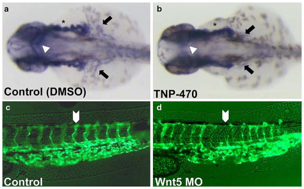
Vascular defects due to chemical inhibition of PCP signaling mimic those of ppt zebrafish. a–b Tg(fli:EGFP) expression (28–30 hpf) as detected by whole mount. Relative to WT (a) vasculature, TNP-470-treated animals (b) displayed disrupted vessel growth (asterisks), disrupted common cardinal vein formation (arrows) and markedly diminished middle cerebral vein (arrowheads). c–d Wnt5 MO-injected embryos evaluated for vasculature defects. Fluorescent imaging of Control (c) and Wnt MO-injected embryos (d), lateral view of the tail. Wnt5-MO injection (d) disrupts intersegmental angiogenesis in Tg(fli:EGFP)