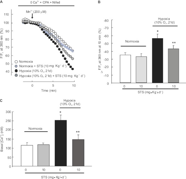
Figure 2.
Effects of STS on store-operated Ca2+ entry (SOCE) and basal intracellular Ca2+ concentration ([Ca2+]i) in pulmonary arterial smooth muscle cells (PASMCs) isolated from rats exposed to hypoxia. PASMCs were isolated from normoxic or hypoxic (10% O2 for 21 d) rats with or without STS (10 mg/kg/d) treatment. (A) SOCE determined by measuring the time course of Fura-2 fluorescence intensity excited at 360 nm before and after adding 200 μM Mn2+ in Ca2+–free Krebs-Ringer bicarbonate solution (0 Ca2+) perfusate containing 10 μM cyclopiazonic acid (CPA) and 5 μM nifedipine (Nifed) in PASMCs. Data at each time point were normalized to fluorescence at time 0 (F/F0). (B) Average quenching of Fura-2 fluorescence by Mn2+. Data are expressed as the percentage decrease in fluorescence at time 10 minutes from time 0. The tested cell numbers in each group were as below: normoxia (115 cells; n = 4), normoxia+STS (117 cells; n = 4), hypoxia (118 cells; n = 4), and hypoxia+STS (117 cells; n = 4). Bar values are mean ± SEM. *P < 0.05 versus normoxic control. **P < 0.05 versus hypoxic control. (C) Basal [Ca2+]i in PASMCs from rats treated with normoxia, normoxia+STS, hypoxia, or hypoxia+STS. Bar values are means ± SEM (n = 5 in each group). *P < 0.05 versus normoxic control. **P < 0.05 versus hypoxic control.

