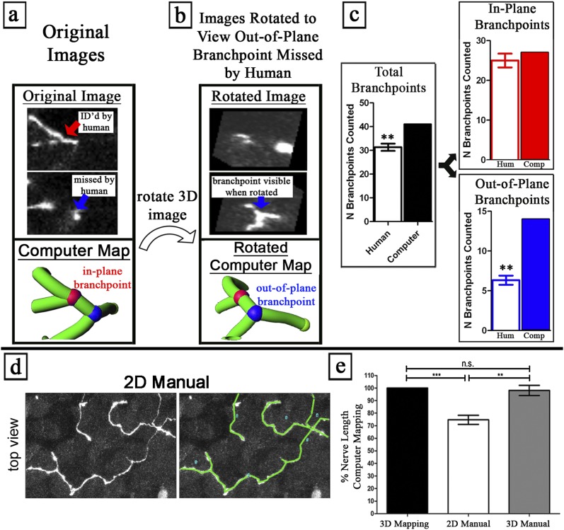
Figure 2.
Computer nerve mapping identifies branchpoints and nerve lengths missed by manual analysis. (a–c) Out-of-plane branchpoints were identified by nerve mapping and not by manual counting. (a) Image slice from nonrotated (“Original Image”) data contains an in-plane branchpoint (red), with three branches viewable in the nonrotated plane. (b) Image slice from rotated data (“Rotated Image”) contains an out-of-plane branchpoint (blue), with branches oriented in a different plane from that in the original images, hence requiring image rotation to visualize. Blinded humans (n = 3) counted branchpoints in the same three-dimensional images. Humans were shown sample in-plane and out-of-plane branchpoints, and were taught to rotate images. (c) Comparison between human branchpoint counting (n = 3) and computer nerve mapping. Humans consistently missed out-of-plane branchpoints that computer nerve mapping correctly identified. See Video 2 in the online supplement. (d–g) A nerve length quantified using computer nerve mapping (“3D Mapping”) is equivalent to the manual quantification of 3D image data (“3D Manual”) and greater than the manual quantification of two-dimensional (2D) flattened image data (“2D Manual”). (d) Manual nerve tracing in two-dimensional and three-dimensional images. (e) Total nerve length was compared with computer mapping. **P < 0.01, ***P < 0.001. n.s., no significance.

