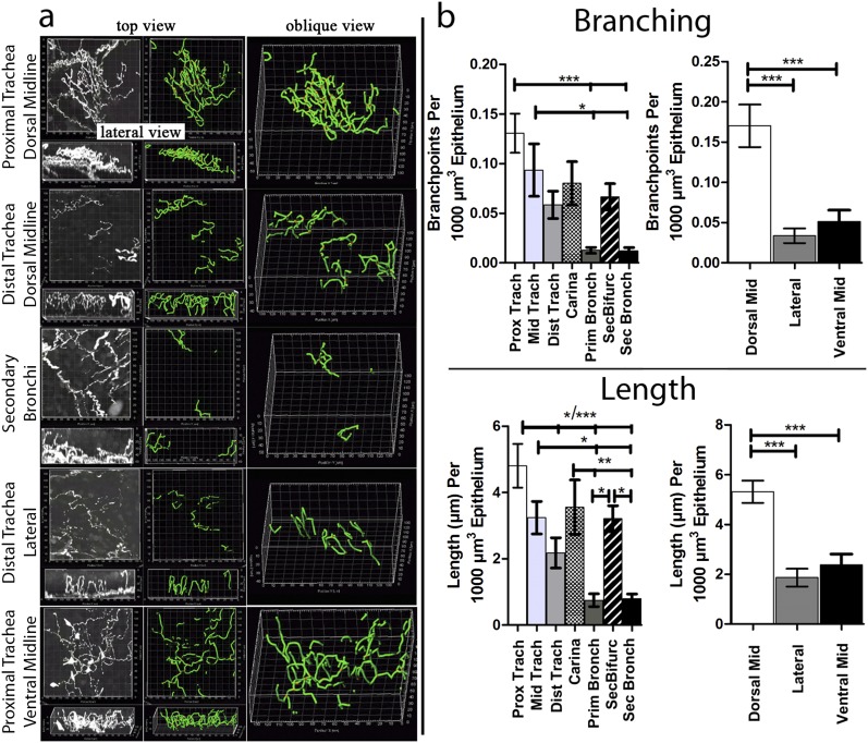
Figure 3.
Organ-wide structural quantification of murine airway epithelial nerves. (a) Top and lateral views of epithelial nerves (white) in different regions of the large airways, with nerve maps (green) overlaid on nerve image data. See Figure E1 for a diagram of airway regions. (b) After mapping, nerve lengths and numbers of branchpoints were quantified by the computer (n = 6). Nerve lengths and branching decreased proximal-to-distal, with regional increases at airway bifurcations. Nerve lengths and branching decreased from the dorsal midline to lateral and ventral positions in the trachea. *P < 0.05, **P < 0.01, ***P < 0.001.

