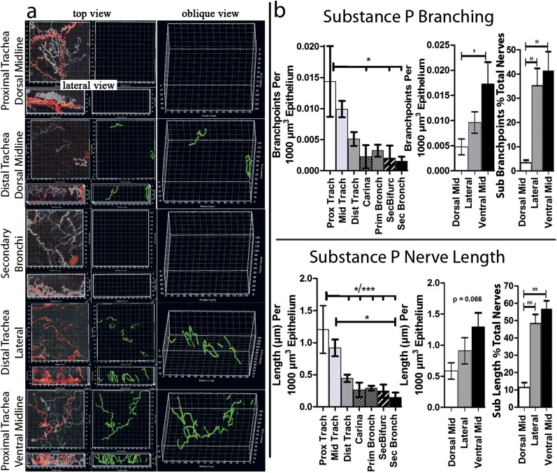
Figure 4.
Distinct organ-wide structure of an airway epithelial nerve subpopulation. (a) Top and lateral views of substance P–expressing nerves (red) were overlaid on nerves labeled with a pan-neuronal marker (white to transparent). Computer substance P nerve map (green) was overlaid on image data. (b) After mapping, epithelial substance P nerve lengths and branching were quantified by the computer (n = 6). Unlike total epithelial nerves identified by PGP 9.5, substance P nerve lengths and branching increased from dorsal to lateral and ventral positions. This incrase was also seen when substance P nerve lengths and branching were calculated as percentages of total nerve lengths and branching. Unlike total nerves, substance P nerve lengths and branching did not increase at airway bifurcations. *P < 0.05, **P < 0.01, ***P < 0.001. See Figure E1 for a diagram of the airway region.

