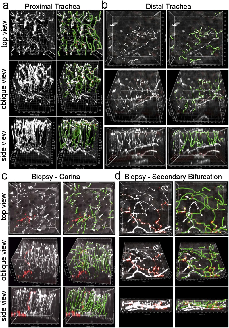
Figure 5.
Epithelial nerve mapping in human airway tissue. (a and b) Top, oblique, and lateral projections of human epithelial nerves (white) in the proximal and distal trachea were overlaid with computer nerve maps (green, with red spheres as branchpoints). (c and d) Epithelial nerves and nerve mapping in airway forceps biopsy tissue from airway bifurcations. Columnar epithelium was present in the trachea autopsy specimens and carina biopsy specimen. Squamous epithelium was present in the secondary bifurcation biopsy specimen. Substance P epithelial nerves (red) are shown in c and d.

