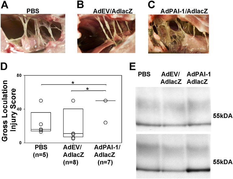
Figure 3.
Overexpression of hPAI-1 markedly increases the severity of tetracycline (TCN)–induced pleural fibrosis. (A–C) The typical level of injury 2 days after intrapleural injection of TCN and 5 days after intrapleural injection of PBS (A), AdEV/AdlacZ (B), or AdPAI-1/AdlacZ (C). (D) The severity of pleural injury in animals injected with PBS (n = 5), empty vector (n = 8), or AdhPAI-1 (n = 7) was evaluated as described briefly in Materials and Methods and detailed in the online supplement, and was expressed as a gross loculation injury for each animal. Data are shown as a box plot in which the 25% and 75% quartiles are indicated by the box and median values are shown as horizontal lines within the box, as previously described (31, 37). Individual scores for each animal are also shown as open circles, which overlap if more than one animal received an identical score. The fibrotic injury observed was uniformly worse (i.e., the gross loculation injury score was higher) in rabbits that received AdPAI-1/AdlacZ compared with PBS (P = 0.029) and AdEV/AdlacZ (P = 0.021). (C) This more complex pleural injury was characterized by the formation of fibrin webs and coalescent fibrinous sheets. (E) Western blot analysis of rabbit plasminogen activator inhibitor–1 (rPAI-1) (top) and hPAI-1 (bottom) antigens in pleural fluids (PFs) of animals with TCN-induced pleural injury. Whereas the concentration of rPAI-1 in the PF of all animals with TCN-induced injury was similar (top), the concentration of hPAI-1 (bottom) was markedly higher in the PF of rabbits transduced with AdPAI-1/lacZ. Monoclonal anti-human PAI-1 antibodies exhibit low-level cross-reactivity with rPAI-1 at higher concentrations.

