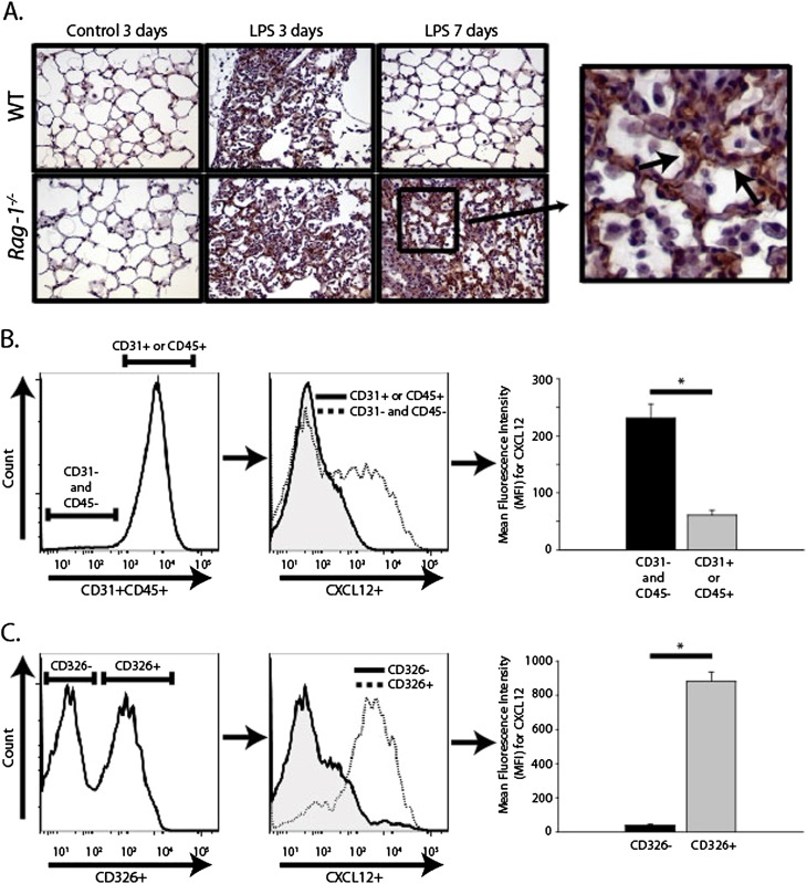
Figure 4.
Epithelial cells were an important source of CXCL12 after LPS-induced lung injury. (A) Lung sections were stained for CXCL12 by immunohistochemistry on Day 3 and Day 7 after intratracheal LPS. Epithelial cells (arrows) appeared to be the predominant source of CXCL12 staining (images are of representative examples, ×200 magnification, n ≥ 2 mice in each group; inset, ×400 magnification). (B) Single-cell suspensions were prepared from the lungs of WT and Rag-1−/− mice on Day 7 after intratracheal LPS or sterile water. Cells were stained with biotinylated anti-CD31 and anti-CD45, followed by V450-conjugated streptavidin as well as allophycocyanin-conjugated (APC)–Cy7–conjugated anti-CD326. Cells were fixed, permeabilized, and stained with APC-conjugated anti-CXCL12. Cells were gated for CD31 and CD45 positivity, and CXCL12 expression was examined by mean fluorescence intensity (MFI) (*P < 0.001, n = 16 mice). (C) CD31−CD45− cells were gated for CD326 (epithelial cell adhesion molecule [EpCAM]) expression to identify epithelial cells. CXCL12 expression in CD326+ and CD326− cells was examined by MFI (*P < 0.001, n = 16 mice).

