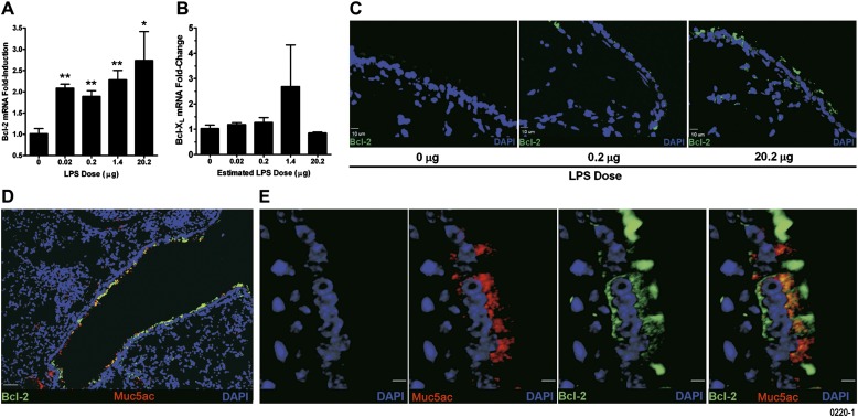
Figure 4.
Aerosolized LPS increases expression of Bcl-2 mRNA and protein in airway epithelial cells. Bcl-2 (A) and Bcl-XL (B) mRNA levels in the lung tissues of mice exposed to LPS as analyzed by quantitative RT-PCR. Data are shown as mean ± SEM (n = 6 per group). *P < 0.05; **P < 0.01. (C) Representative micrographs of lungs from mice exposed to 0.2 (middle panel) and 20.2 μg (right panel) compared with 0 μg (left panel) LPS showing airway epithelial cells that are Bcl-2 immunopositive (green). 4',6-Diamidino-2-phenylindole (DAPI) was used as nuclear counter stain and is shown in blue (scale bar, 10 μM). (D) A representative photomicrograph of a lung section from mice exposed to 20.2 μg of aerosolized LPS showing Bcl-2 and Muc5ac coexpression in airway epithelial cells (scale bar, 50 μM). (E) High-magnification images of Bcl-2– and Muc5ac-immunostained lung sections from mice exposed to 20.2 μg aerosolized LPS. The panels show DAPI-stained nuclei (blue), Muc5ac-positive (red), and Bcl-2–positive (green) cells. The composite image shows colocalization of Muc5ac and Bcl-2 (yellow) (scale bar, 5 μM).

