Figure 5.
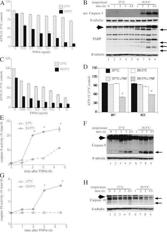
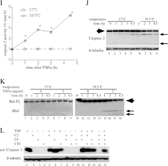
Concurrent exposure to febrile-range hyperthermia (FRH) (39.5°C) augments extrinsic apoptosis through NF-κB– and HSF1-independent pathways. (A) MLE15 cells were incubated for 24 hours at 37°C or 39.5°C with the indicated doses of TNF-α and cell survival was measured by crystal violet staining. (B) MLE15 cells were treated with 2 ng/ml TNF-α at 37°C or 39.5°C for the indicated time, and cells were lysed and immunoblotted for active caspase-3, PARP, and β-tubulin. (C) IκBSR-expressing MLE15 cells were treated as described in (A) and cell survival assessed by crystal violet staining. (D) WT and HSF1-KO MEFs were incubated for 4 hours at 37°C or 39.5°C with or without 4 ng/ml TNF-α and cell survival was assessed by crystal violet staining. (E–K) MLE15 cells were incubated with 2 ng/ml TNF-α at 37°C or 39.5°C and sequentially lysed for fluorimetric (E, G, and I) and immunoblot (F, H, and J) analysis of caspase-8 (E and F), caspase-10 (G and H), and caspase-2 (I and J), and for immunoblot analysis of Bid (K). (F, H, J, and K) Caspase and Bid cleavage products are indicated by arrows. Full-length Bid and caspases are indicated by large arrowhead. All graphs show means (±SE) of four experiments; *P < 0.05 versus 37°C. (A and C) There was a difference between 37°C and 39.5°C and between TNF-α–treated and untreated at 37°C (P < 0.0001 by MANOVA). (F, H, I, J, and K) Each panel is representative of four similar blots. (L) MLE15 cells were pretreated with inhibitors of caspase-2, -8, and -10 (50 nM) alone or in combination for 40 minutes at 37°C, then were incubated at 39.5° with or without 2 ng/ml TNF-α and immunoblotted for activated caspase-3. A representative of three similar blots is shown.


