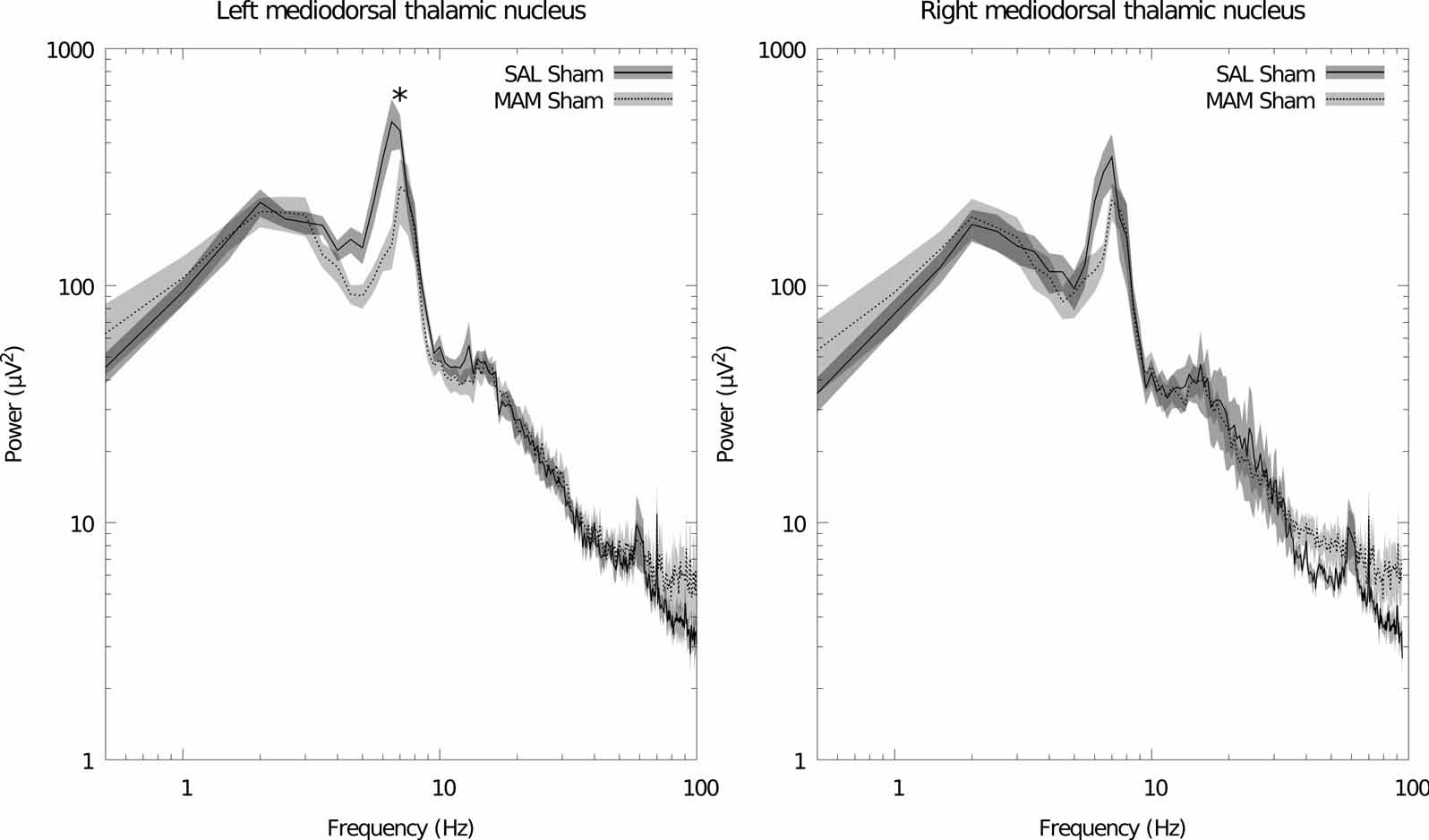
Figure 1.
MAM treatment induces a deficit in mediodorsal thalamic nucleus theta power. This deficit is not restored by ventral hippocampal stimulation (data not shown). Left: grand averaged power spectra for the left hemisphere. Right: grand averaged power spectra for the right hemisphere. Data is plotted as the mean (solid/dashed line) ± SEM (shaded region). * indicates a significant difference between sham-stimulated SAL and MAM treated animals for that frequency bin.

