
Figure 5. Diamide enhances thapsigargin-mediated changes in [Ca2+]i of HAECs.
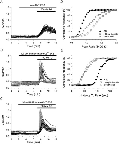
Fura-2-loaded HAECs in zero Ca2+-ECS were left untreated (A), or treated with either 30 nm HIST (C) or with 100 μm diamide (B) for 5 min immediately prior to challenging with 300 nm thapsigargin (TG). A–C, cells were segregated into two phenotypic profiles based on both the rate and the magnitude of the TG-mediated change in [Ca2+]i. Cells represented by black lines displayed a slow rate of rise and a small peak change in [Ca2+]i, whereas cells represented by greyscale lines exhibited an enhanced response characterized by a rapid rate of rise and a large change in [Ca2+]i. D, cumulative frequency of the maximal change in fura-2 fluorescence (peak ratio) observed in response to TG in control (filled circles), diamide (open squares) and HIST (grey triangles) treated cells. E, cumulative frequency of the time from TG exposure to the maximal change in fura-2 fluorescence (latency to peak) for control (filled circles), diamide (open squares), and HIST (grey triangles) treated cells. A total of 121 (control), 135 (diamide) and 149 (HIST) cells were analysed from 4 experiments for each experimental condition. D and E, the TG-induced peak ratio and latency to peak for both diamide- and HIST-treated cells were significantly different from untreated controls; P < 0.001.