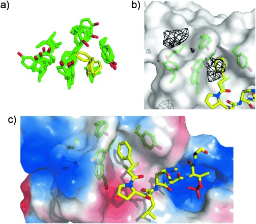
Figure 4.

Design of a chimeric peptide ligand based on an alternative binding mode obtained from ligand-mapping simulations. a) Superposition of five snapshots of PBD binding pocket (green) from a ligand-mapping MD trajectory, showing an alternative binding mode of benzene (yellow). b) Binding mode of chimeric peptide (yellow sticks) at PBD pocket (green sticks) overlaid with benzene probability isosurfaces (black mesh) for the protein structure shown. c) Crystal structure of PBD complexed with chimeric peptide (yellow sticks). Regions of positive and negative electrostatic potential on the PBD surface are colored blue and red, respectively.
