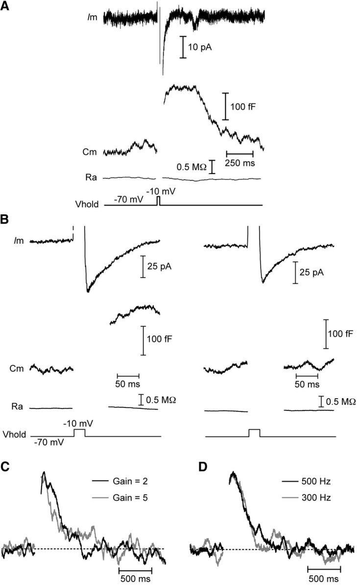
Figure 2.

Capacitance responses are not conductance or amplifier artifacts. Whole-cell recordings from cones in salamander retinal slice. A, B, Capacitance changes are independent of the calcium-activated chloride tail current, ICl(Ca). A, A capacitance change (Cm) was triggered by a 25 ms step depolarization from −70 to −10 mV when ICl(Ca) was completely blocked by niflumic acid (200 μm) in the superfusate. Baseline values: Cm = 41.2 pF, Ra = 50.0 MΩ. B, In the absence of niflumic acid, a 25 ms depolarizing pulse evoked a large ICl(Ca) and change in capacitance (left). Following a 1 s pulse train (25 ms steps to −10 mV at 13.3 Hz) to empty the releasable pool of vesicles, another depolarizing pulse did not evoke an exocytotic capacitance change despite the presence of a prominent ICl(Ca) (right). Baseline Cm = 48.2 pF, Ra = 38.1 MΩ. C, D, Normalized capacitance responses showing that the time course of endocytosis was unaffected when the gain of the amplifier was switched from 2 to 5 (C) or when the frequency of the sine wave used for capacitance measurements was decreased to 300 Hz from 500 Hz (D). Vhold, holding potential; Ra, access resistance.
